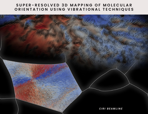
A groundbreaking study that allows the creation of 3D maps of bond orientations in a sample has been published by the CIRI beamline team in the Journal of American Chemical Society. In-depth characterisation of a sample, while not destroying the research material, is crucial for scientists in material and life sciences and will allow previously unattainable information to be extracted from complex systems.
If we are able to obtain a thermal conductor from a common insulator by changing the orientation of the particles, isn't the study of orientation and spatial visualisation of macromolecules crucial information for understanding each system? That's the question the NCPS SOLARIS researchers asked themselves. By using known research methods and applying custom mathematical analysis to experimental data, they were able to push the boundaries of the technique and obtain high resolution images visualising the spatial orientation of the molecules in the spherulite polymer under study.
Infrared (IR) spectroscopy allows obtaining rich information about the studied sample without destroying it or using sample staining. The technique has been successfully used in many scientific fields for years, and it is crucial in areas of new materials research. Scientists at the SOLARIS Synchrotron, led by Dr hab. Tomasz Wróbel,, applied the so-called Concurrent Analysis (4P-3D) to infrared spectromicroscopic data for the first time. With this, they obtained information on the orientation angles of macromolecules in a sample of polycaprolactone spherulite. This turned out to be possible by simultaneously analysing two bands with roughly perpendicular transition moment orientations measured at 4 different linear polarisations.
'The 3D molecule orientation in a material, next to its chemical composition, is the basic information we want to obtain about the sample', says Dr hab. Tomasz Wróbel (SOLARIS). 'It affects the mechanical properties (tensile, fracture), chemical properties, physiochemistry of the surface, conductivity, diffusion of, for example, a drug through the tissue just to name a few. It's a whole bunch of different sample properties, and we need tools to study and determine them'.
Until now, it has been possible to study the orientation of bonds in the sample, but only to a certain extent, e.g. over large areas, in thin layers or by using tomography. With the latest discovery, it is possible to obtain high-resolution 3D images and, importantly, without destroying the material under study and without using additional dyes or labels.
'Furthermore, we show that the method can be applied to high-resolution (diffraction-limited) FT-IR and Raman imaging, and even to super-resolution O-PTIR imaging', highlights Paulina Kozioł (CIRI, SOLARIS), the first author of the publication. The O-PTIR measurements were made possible at the SOLEIL synchrotron thanks to a collaboration with Dr. Ferenc Borondics.
Spatial, non-destructive orientation studies will have a profound impact on materials and life sciences as a method for extracting previously unattainable information from complex systems at the nanometer scale. We already know that the performance of solar panels depends on the orientation and order of the polymers inside. Such fundamental research brings us closer to more efficient and environmentally friendly engineering solutions.
Link to publication - https://pubs.acs.org/doi/full/10.1021/jacs.2c05306
The research was funded by an NCN Sonata grant No 2018/31/D/ST4/01833.
Source: Official press release