On 25 October 2018, the Polish Ministry of Science and Higher Education has officially announced its decision to fund a unique cryogenic electron microscope (Cryo-EM) and establish the National Centre for Cryogenic Electron Microscopy at the JU National Synchrotron Radiation Centre SOLARIS. It’s a direct result of lengthy organisational work and unwavering support of 17 research institutions, including the Jagiellonian University, which have urged the Ministry to fund such an endeavour.
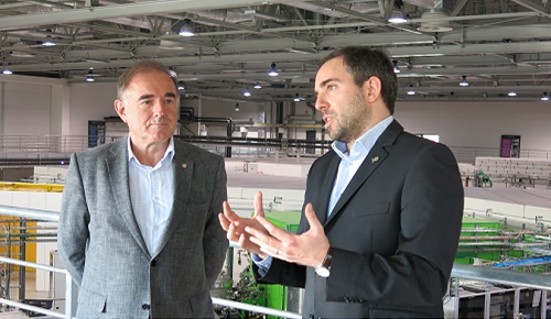
We asked Prof. Marek Stankiewicz, Director of the SOLARIS Centre, and Dr Sebastian Glatt from the JU Małopolska Centre of Biotechnology to explain the benefits of possessing such a device in Kraków.
‘It’s a wonderful example of collaboration of the Polish academic community. Scientists from various research centres came together in order to voice their support for a great idea’, said Prof. Stankiewicz. The microscope will be available to be used by any institution, providing an opportunity for further integration of Polish and European researchers. Although it won’t start operating until the second half of the next year, 31 research teams have already expressed their willingness to use it in their work. ‘We treat this project as an enormous investment in the Polish scientific community. It’s really an amazing gift for our research teams in the field of molecular biology’, stated Dr Glatt. The machine will be under a constant supervision of a dedicated, highly trained specialist – a testament to its complexity. Aside from making sure the microscope is properly maintained, the specialist will guarantee the reliability of any data that might be acquired through its use.
So what’s it all about? Why does the Cryo-EM technology act like a magnet for scientists? What are the practical aspects of the measurements carried out by this particular microscope?
Doors wide open
Humanity’s first attempts to observe the fundamental elements that constitute matter – particles and atoms – might be compared to looking through a keyhole. There wasn’t a whole lot to look at, despite additional input from the observation of electromagnetic spectrum (think of it as atoms knocking on the door from the other side). It was quite a daunting task. Developments in particle observation came at a slow pace: first with the improvement of the optical microscope, and then with the invention of the scanning electron microscope (SEM) and fluorescence microscope. Step by step, the door was set ajar. Now, with Cryo-EM technology, we’ve opened this door wider than ever before and look at matter at the atomic level. It’s never been that easy.
Enter cryogenics
What exactly is the difference between cryogenic microscopes and the other advanced methods used beforehand? The first and rather obvious difference is that Cryo-EM makes use of cryogenic-level temperature (i.e. extremely low – about -150°C). This is the temperature of water in which samples are frozen before they’re tested, which is already a great help. It stabilises the samples, making them much more stable – an important quality, considering their size is usually measured in micrometres.
To be sure, freezing water doesn’t seem like a game-changer in and of itself. But it isn’t about if the water is frozen; it’s about how it’s frozen. We need to remember that the process of freezing turns water into ice with a regular crystal structure. Instead of travelling straight through the ice and hitting the sample, the beam emitted by an ordinary electron microscope bounces off of crystal surfaces, leading to in erroneous measurement results.
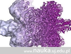
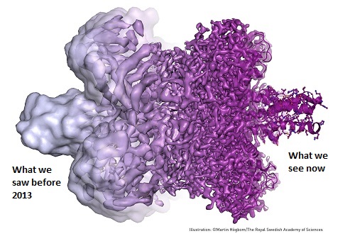 The whole point is, therefore, to freeze water in such a way that it doesn’t produce the regular crystal structure, but rather becomes amorphous. Not only would that make it easier for the beam to pierce through it, but also preserve samples from suffering any damage. Jacques Dubochet, one of the winners of the 2017 Nobel Prize in Chemistry, was awarded for resolving this issue. He invented a way of cryogenically freezing water containing microscope samples in just a few nanoseconds. Water particles are unable to arrange themselves in an orderly fashion and become locked in an amorphous solid form, safe for samples and suitable for studying. Now we must add another element: an innovative technique that employs a lower-energy electron beam, developed by Richard Henderson, who was also awarded with the 2017 Nobel Prize in Chemistry. It delivers extremely high resolution images of the samples' natural atomic environment, providing up to 13 TB (!) of data per day. But therein lies another problem: it requires an immense computing power to turn this many 2D images into 3D models. Joachim Frank, the third winner of the 2017 Nobel Prize in Chemistry mentioned in this article, came up with a way to solve it. He developed highly advanced methods of mathematical image processing, contributing to obtaining 3D models of molecules of phenomenal quality.
The work of the three abovementioned scientists resulted in a cutting-edge device, the Cryo-EM microscope, which introduced us into a whole new world of research.
Breakthroughs
What branches of science can benefit from a cryogenic electron microscope? Already it’s widely considered a revolution in structural biology. ‘The microscope brings us closer to understanding the fundamentals of protein complexes, basics of DNA repair, and new ways of fighting cancer, Alzheimer’s, and obesity’, explained Dr Glatt. ‘The human organism, the way it fights off bacteria and reacts to harmful conditions – it’s all based on molecular mechanisms. The microscope holds a great potential for improving our health’, added Prof. Stankiewicz.
We’re standing at the precipice of a vast sea of possibilities for Polish scientists. There will be opportunities of international collaboration and world-class research projects fuelled by ultraprecise measurements provided by the microscope. But what’s more, there is also hope for new methods of treatment that will protect what we value the most: the human life.
Image: Royal Swedish Academy of Science
Original text: www.nauka.uj.edu.pl